Instytut Podstawowych Problemów Techniki
Polskiej Akademii Nauk

Partnerzy

W. Dobkowska-Chudon

District Hospital (PL)

Ostatnie publikacje
1.  Dobkowska-Chudon W., Wróbel M., Frankowska E., Zegadło A., Krupniewicz A., Nowicki A., Olszewski R., Comparison of acoustocerebrography measurement and magnetic resonance imaging methods in the assessment of white matter lesions in patients with atrial fibrillation, ARCHIVES OF ACOUSTICS, ISSN: 0137-5075, DOI: 10.24425/aoa.2020.134060, Vol.45, No.3, pp.445-452, 2020

Streszczenie:
The brain is subject to damage, due to ageing, physiological processes and/or disease. Some of the damage is acute in nature, such as strokes; some is more subtle, like white matter lesions. White matter lesions or hyperintensities (WMH) can be one of the first signs of micro brain damage. We implemented the Acoustocerebrography (ACG) as an easy to use method designed to capture differing states of human brain tissue and the respective changes. Aim: The purpose of the study is to compare the efficacy of ACG and Magnetic Resonance Imaging (MRI) to detect WMH in patients with clinically silent atrial fibrillation (AF). Methods and results: The study included 97 patients (age 66.26 ± 6.54 years) with AF. CHA2DS2- VASc score (2.5 ±1.3) and HAS BLED (1.65 ± 0.9). According to MRI data, the patients were assigned into four groups depending on the number of lesions: L0 – 0 to 4 lesions, L5 – 5 to 9 lesions, L10 – 10 to 29 lesions, and L30 – 30 or more lesions. Authors found that the ACG method clearly differentiates the groups L0 (with 0–4 lesions) and L30 (with more than 30 lesions) of WMH patients. Fisher's Exact Test shows that this correlation is highly significant (p < 0.001). Conclusion: ACG is a new, easy and cost-effective method for detecting WMH in patients with atrial fibrillation. The ACG measurement methodology should become increasingly useful for the assessment of WMH.

Słowa kluczowe:
acoustocerebrography, brain MRI, atrial fibrillation, white matter hyperintensities

Afiliacje autorów:
Dobkowska-Chudon W. - District Hospital (PL)
Wróbel M. - Fraunhofer Institute for Cell Therapy and Immunology IZI (DE)
Frankowska E. - Military Medical Institute (PL)
Zegadło A. - inna afiliacja
Krupniewicz A. - Medical University of Warsaw (PL)
Nowicki A. - IPPT PAN
Olszewski R. - IPPT PAN
70p.
2.  Dobkowska-Chudon W., Wrobel M., Karłowicz P., Dabrowski A., Krupienicz A., Targowski T., Nowicki A., Olszewski R., Detecting cerebrovascular changes in the brain caused by hypertension in atrial fibrillation group using acoustocerebrography, PLOS ONE, ISSN: 1932-6203, DOI: 10.1371/journal.pone.0199999, Vol.13, No.7, pp.1-10, 2018

Streszczenie:
Acoustocerebrography is a novel, non-invasive, transcranial ultrasonic diagnostic method based on the transmission of multispectral ultrasound signals propagating through the brain tissue. Dedicated signal processing enables the estimation of absorption coefficient, frequency-dependent attenuation, speed of sound and tissue elasticity. Hypertension and atrial fibrillation are well known factors correlated with white matter lesions, intracerebral hemorrhage and cryptogenic stroke numbers. The aim of this study was to compare the acoustocerebrography signal in the brains of asymptomatic atrial fibrillation patients with and without hypertension. The study included 97 asymptomatic patients (40 female and 57 male, age 66.26 +/- 6.54 years) who were clinically monitored for atrial fibrillation. The patients were divided into two groups: group I (patients with hypertension) n = 75, and group II (patients without hypertension) n = 22. Phase and amplitude of all spectral components for the received signals from the brain path were extracted and compared to the phase and amplitude of the transmitted pulse. Next, the time of flight and the attenuation of each frequency component were calculated. Additionally, a fast Fourier transformation was performed and its features were extracted. After introducing a machine learning technique, the ROC plot of differentiations between group I and group II with an AUC of 0.958 (sensitivity 0.99 and specificity 0.968) was obtained. It can be assumed that the significant difference in the acoustocerebrography signals in patients with hypertension is due to changes in the brain tissue, and it allows for the differentiating of high-risk patients with asymptomatic atrial fibrillation and hypertension.

Słowa kluczowe:
changes in the brain, hypertension in atrial, acoustocerebrography

Afiliacje autorów:
Dobkowska-Chudon W. - District Hospital (PL)
Wrobel M. - Sonovum A.G. (DE)
Karłowicz P. - Sonomed Sp. z o.o. (PL)
Dabrowski A. - MTZ Clinical Research (PL)
Krupienicz A. - Medical University of Warsaw (PL)
Targowski T. - National Institute of Geriatrics, Rheumatology and Rehabilitation (PL)
Nowicki A. - IPPT PAN
Olszewski R. - IPPT PAN
40p.

Abstrakty konferencyjne
1.  Olszewski R., Dobkowska-Chudon W., Wrobel M., Karlowicz P., Dabrowski A., Krupienicz A., Targowski T., Nowicki A., Is Acoustocerebrography a new noninvasive method for early detection of the brain changes in patients with hypertension?, ESC Congress 2017, European Society of Cardiology Congress 2017, 26-30 August, Barcelona, Spain, 2017-08-26/08-30, Barcelona (ES), DOI: 10.1093/eurheartj/ehx501.P190, Vol.38, No.suppl_1, pp.36, 2017

Streszczenie:
Background: Hypertension (HT) is the leading cause of global disease burden and overall health loss. The brain is one of the main target organs affected by HT. HT is a potentially modifiable risk factor that leads to the formation of large vessel macroangiopathy, small vessel disease, microangiopathy, and microhemorrhages. Early detection of the brain changes (BC) gives a chance to receive appropriate treatment and protection from irreversible damage. Acoustocerebrography (ACG) is a set of techniques to capture the states of human brain tissue, and its changes on its molecular and cellular level. It is based on noninvasive measurements of various parameters obtained by analyzing an ultrasound pulse emitted across the human's skull. The main idea of this method relies in the relation between the tissue density, bulk modulus, and speed of propagation, for ultrasound waves in this medium. In our previous studies we showed that ACG is an effective method for detecting white matter lesions compared to the Magnetic Resonance Imaging. Additionally we showed that ACG allows to obtain a differentiated signal originates from atrial fibrillation (AF) patients and high-risk patients wit AF and HT.
Aim: The aim of the study was early detection of the BC in patients with HT using ACG.
Methods: The study included 136 female and 98 male patients (age 43.6±15.7 years) who were surveyed in the clinical research. The patients were divided into two groups: group I (patients with HT) n=33, and control group II (patients without HT) n=201. Phase and amplitude of all frequency components of the received signals from the brain path were extracted and compared to the phase and amplitude of the transmitted pulse. By doing so, the time of flight and the attenuation of each frequency component were calculated. Additionally, a fast Fourier transformation (FFT) was performed and its features were extracted.
Results: After introducing a machine learning technique, the ROC plot with an AUC of 0.929 with sensitivity 0.879 and specificity 0.831 was obtained (Fig. 1).
Conclusion: ACG is new promising method, which allows for early detection of change in the brain in the patients with HT.

Afiliacje autorów:
Olszewski R. - IPPT PAN
Dobkowska-Chudon W. - District Hospital (PL)
Wrobel M. - Sonovum A.G. (DE)
Karlowicz P. - Sonomed Sp. z o.o. (PL)
Dabrowski A. - MTZ Clinical Research (PL)
Krupienicz A. - Medical University of Warsaw (PL)
Targowski T. - National Institute of Geriatrics, Rheumatology and Rehabilitation (PL)
Nowicki A. - IPPT PAN

Kategoria A Plus

IPPT PAN

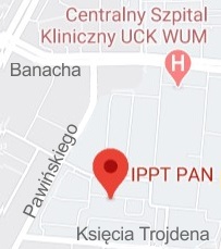
logo ippt            ul. Pawińskiego 5B, 02-106 Warszawa
  +48 22 826 12 81 (centrala)
  +48 22 826 98 15
 

Znajdź nas

mapka
© Instytut Podstawowych Problemów Techniki Polskiej Akademii Nauk 2024